Scleral lenses have emerged as the most effective treatment for keratoconus, but their effectiveness hinges on meticulous design and fitting. Today’s scleral lenses can be made to fit better than ever before thanks to new ocular imaging technologies. This means that they are safer, more comfortable, and more effective at correcting vision than ever .
In this blog post, we’ll talk about how imaging tools like anterior segment OCT, corneal topography, and scleral profilometry are changing the way specialty lenses are used and how they are helping keratoconus patients in 2025.
Why it's important to be precise when fitting scleral lenses
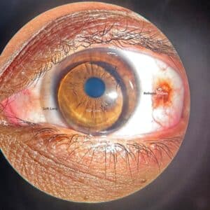
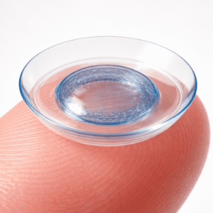
Scleral lenses don’t sit on the cornea like soft lenses do. Instead, they sit on the sclera and vault over the cornea. Understanding the three-dimensional shape of the eye is crucial for this process.
If scleral lenses don’t fit well, they can cause:
- Lens discomfort or intolerance
- They may touch the cornea or cause mechanical damage.
- Conjunctival blanching and impingement
- Fogging and collecting debris
Patients experience less time spent wearing lenses and report lower satisfaction.
Advanced imaging eliminates the need for guesswork. It lets doctors make lenses that fit the patient’s eye anatomy perfectly, which is essential for keratoconus because every eye is different.
The Imaging Technologies That Make Modern Lens Design Possible
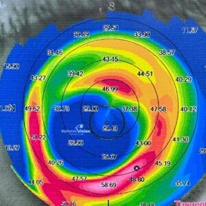
1. Topography of the cornea
This technique shows the curvature of the front surface of the cornea, pointing out steep areas, cones, and other problems. It’s the first step in figuring out if someone has keratoconus and planning the lens parameters.
This aids in determining the location and severity of the cone.
This aids in determining the starting lens diameter and sagittal depth.
Also helpful for designing soft and hybrid lenses.
2. Optical Coherence Tomography of the Anterior Segment (AS-OCT)
OCT makes obvious cross-sectional pictures of the cornea, limbus, and sclera.
Measures vault over the cornea to make sure it doesn’t touch it.
It also measures the thickness of the tear reservoir located behind the lens.
It helps identify edge lift or compression that may occur after wearing the lenses.
Let doctors make small changes to the design of the lens.
OCT is also important for follow-ups to make sure the eyes are still healthy.
3. Scleral Profilometry (for example, EyePrintPRO, sMap3D, and Pentacam CSP)
This is a new tool that scans the whole front of the eye, including the sclera.
Maps the sclera’s irregularities, toricity, and differences in elevation.
It is necessary to make scleral lenses that are freeform or in a specific quadrant.
Very helpful for advanced keratoconus, corneas that have had surgery, or asymmetry in the conjunctiva.
Scleral profilometry is making lenses much more accurate and cutting down on the number of trial fittings by a lot.
4. Aberrometry of the wavefront
Some high-tech clinics use aberrometry to look at high-order aberrations and make
wavefront-guided scleral lenses that give you better vision than regular lenses.
How Imaging Helps People with Keratoconus
Higher first-fit success rate: fewer trial lenses and faster fitting.
Better alignment means more comfort and longer wear time.
Lenses are made to fit the shape of each eye, so vision is stable and sharper.
Identifying issues early, such as excessive corneal clearance or lens impingement, is crucial.
Customization: Lenses can be asymmetric or freeform as needed
An Example of a Clinical Case
A young patient with advanced keratoconus and a steepening inferior cone couldn’t wear RGPs. We used AS-OCT and profilometry to make a toric scleral lens with a higher vault on the bottom. The patient had 20/25 vision and could wear the contact lenses comfortably for 12 hours a day without any signs of hypoxia or corneal touch.
What Patients Need to Know Before Getting Fitted
Your doctor may use more than one tool to check your eyes.
Digital scans can take just a few seconds but give a lot of information.
These tools help you fit better and spend less time in the chair.
If you have them, bring your old lenses or records.
Conclusion
In 2025, imaging has turned the art of fitting scleral lenses into a science. Tools like AS-OCT, corneal topography, and profilometry have made it much easier for us to treat keratoconus with custom, comfortable, and effective scleral lenses. If you’ve had lens issues or are considering surgery, it’s time to explore imaging-guided lens fitting.
About the Writer
Reform Vision Hyderabad’s Reekham Lal is an expert in designing scleral lenses and fitting them using imaging. He is an expert in difficult keratoconus, corneas after surgery, and ocular surface disease, and he uses the latest technology to get great results.