1. Patient Information
● Name: [Confidential]
● Age/Sex: 32 years / Female
● Occupation: Teacher
● Referred by: Retina Specialist for Low Vision Evaluation
● Chief Complaint: Progressive reduction in vision since teenage years, difficulty in reading, mobility issues in dim light, and glare sensitivity.
2. History
● Ocular History:
○ Diagnosed with Retinitis Pigmentosa (RP) at age 16.
○ High myopia since childhood (spectacle correction around –12.00 D OU).
○ Reports night blindness and peripheral field loss worsening over last 5 years.
○ No previous low vision rehabilitation.
● Systemic History: Non-contributory.
● Family History: Positive for RP in maternal uncle (suggestive of hereditary RP).
● Medications: None currently.
3. Clinical Examination
3.1 Visual Acuity (with best correction)
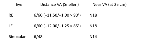
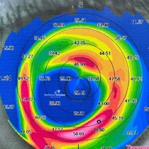
3.2 Refraction
High myopic astigmatism in both eyes, no significant improvement with further correction.
4. Functional Vision Assessment
● Contrast Sensitivity: Reduced (Pelli–Robson: 0.90 log units OU)
● Color Vision: Impaired blue–yellow axis (Farnsworth D-15 test – tritan defect pattern)
● Glare Sensitivity: Severe discomfort under bright light (photophobia)
● Fixation Stability (Macular Integrity): Central fixation preserved
● Binocularity: No suppression; limited fusion due to field constriction
5. Visual Field Assessment (Humphrey Visual Field – 30-2 Program)
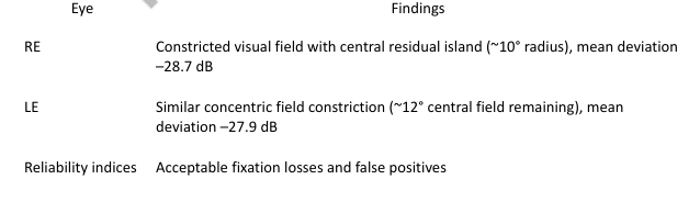
Interpretation: Severe peripheral field loss typical of advanced retinitis pigmentosa; functional central field retained for reading and near tasks.
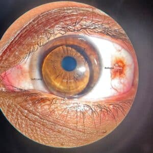
6. Retinal Investigations
● Fundus Examination (Optos Ultra-widefield):
○ Attenuated retinal vessels, waxy disc pallor, and bone-spicule pigmentation in mid-periphery.
○ Myopic fundus with posterior staphyloma.
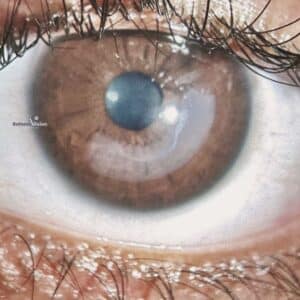
● Optical Coherence Tomography (OCT):
○ Thinning of outer retinal layers, preserved foveal contour, and reduced ellipsoid zone integrity.
● Fundus Autofluorescence (FAF):
○ Hyperautofluorescent ring around the macula suggestive of viable photoreceptor zones.
● Electroretinogram (ERG):
○ Severely reduced scotopic and photopic responses confirming advanced RP.
7. Low Vision Assessment and Intervention
7.1 Distance Vision Enhancement
● Objective: To optimize remaining central vision for mobility and television viewing.
● Trialed Devices:
○ 3× Galilean telescope (mounted monocularly on RE) — improved recognition of 2 m signage.
○ 2.5× binocular telescope with lightweight frame for intermediate distance tasks (classroom board reading).
● Final Prescription: 3× Galilean monocular telescope for distance spotting.
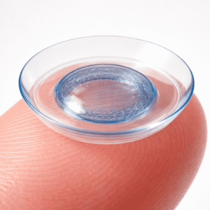
7.2 Near Vision Enhancement
● Objective: Improve reading and near task performance using residual central vision.
● Trialed Devices:
○ 4× illuminated hand magnifier – achieved N8 continuous reading.
○ 3× dome magnifier – preferred for stability and ease of use.
○ CCTV (video magnifier) evaluation – effective for prolonged reading with contrast and reverse polarity modes.
● Final Prescription: 4× LED-illuminated hand magnifier and recommendation for home-based electronic magnifier use.
7.3 Non-optical Interventions
● Use of anti-glare amber filters (450 nm cut-off) to enhance contrast and reduce photophobia.
● Task lighting: Directed LED lamp with adjustable brightness.
● Environmental modification: High-contrast labels and large-print materials.
7.4 Orientation and Mobility Training
● Counseled for mobility cane use in dim light.
● Suggested contrast markings on home stairs and furniture edges.
● Referred for orientation and mobility training at a rehabilitation center.
7.5 Counseling and Rehabilitation
● Explained prognosis of RP and potential for future assistive technology (AT) integration — such as portable digital magnifiers and voice-based reading apps.
● Discussed low vision support group enrollment for psychological support.